How to Prepare

On the day of the exam you may eat normally. You should not take calcium supplements for at least 24 hours before your exam.
You should wear loose, comfortable clothing, avoiding garments that have zippers, belts or buttons made of metal. Objects such as keys or wallets that would be in the area being scanned should be removed.
You may be asked to remove some or all of your clothes and to wear a gown during the exam. You may also be asked to remove jewelry, removable dental appliances, eye glasses and any metal objects or clothing that might interfere with the x-ray images.
Inform your physician if you recently had a barium examination or have been injected with a contrast material for a computed tomography (CT) scan or radioisotope scan. You may have to wait 10 to 14 days before undergoing a DXA test.
Women should always inform their physician and x-ray technologist if there is any possibility that they are pregnant. Many imaging tests are not performed during pregnancy so as not to expose the fetus to radiation. If an x-ray is necessary, precautions will be taken to minimize radiation exposure to the baby. See the Safety page for more information about pregnancy and x-rays.

Dexa

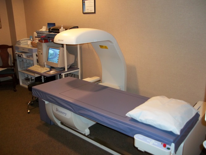
Bone Density Testing (DEXA)

DEXA stands for 'dual energy X-ray absorptiometry'. It is a test that measures the density of bones. Density means how much of something there is in a certain amount of space. The denser the tissue, the less X-rays pass through. Air and water are less dense than solid things such as bone. This is because the particles which make air and water are not held closely together. In general, the more dense the bone, the stronger it is, and the less likely it is to break.

100 0246
Patients must alert the technologist if they may be pregnant.


Who should have a DEXA scan?

A DEXA scan may be advised if you are at increased risk of osteoporosis. Osteoporosis usually causes no symptoms at first. However, if you have osteoporosis, you have an increased risk of breaking a bone. (See separate leaflet called Osteoporosis for more details.) If a DEXA scan shows that you have osteoporosis, then you may be given advice and treatment to help strengthen your bones. Therefore, a DEXA scan may be advised if you have:

  • A fracture following a minor fall or injury.

  • Loss of height due to fracture of a vertebra (back bone).

  • Taken steroid tablets for three months or more.

  • An early menopause (aged less than 45).

  • A history of periods stopping (amenorrhoea) for more than one year before the menopause.

  • Other disorders associated with osteoporosis such as rheumatoid arthritis or coeliac disease.

  • A family history of hip fracture on your mother's side of your family.

  • A body mass index of less than 19 (that is, if you are very underweight.)


How Does It Work?

A DEXA scan uses low-energy X-rays. A machine sends X-rays from two different sources through the bone being tested. Bone blocks a certain amount of the X-rays. The more dense the bone is, the less X-rays get through to the detector. By using two different X-ray sources rather than one it greatly improves the accuracy in measuring the bone density.

The amount of X-rays that comes through the bone from each of the two X-ray sources is measured by a detector. This information is sent to a computer which calculates a score of the average density of the bone. A low score indicates that the bone is less dense than it should be, some material of the bone has been lost, and it is more prone to fracture.

DEXA Scan

A DEXA scan is a test designed to measure bone mineral density and diagnose osteoporosis. This test is quick and painless. After initial screening, DEXA's are done every 3-5 years or every 2-3 years if osteoporosis or osteopenia is detected. In the past, osteoporosis could be detected only after you broke a bone. By that time, however, your bones could be quite weak. A bone density test enhances the accuracy of calculating your risk of breaking bones. A bone density test uses X-rays to measure how many grams of calcium and other bone minerals are packed into a segment of bone. The bones that are most commonly tested are in the spine, hip and forearm.