How is it Performed?

MRI examinations may be performed on outpatients or inpatients.

You will be positioned on the moveable examination table. Straps and bolsters may be used to help you stay still and maintain the correct position during imaging.

Devices that contain coils capable of sending and receiving radio waves may be placed around or adjacent to the area of the body being studied.

If a contrast material will be used in the MRI exam, a physician, nurse or technologist will insert an intravenous (IV) catheter, also known as an IV line, into a vein in your hand or arm. A saline solution may be used. The solution will drip through the IV to prevent blockage of the IV catheter until the contrast material is injected.

You will be moved into the magnet of the MRI unit and the radiologist and technologist will perform the examination while working at a computer outside of the room.

If a contrast material is used during the examination, it will be injected into the intravenous line (IV) after an initial series of scans. Additional series of images will be taken during or following the injection.

When the examination is completed, you may be asked to wait until the technologist or radiologist checks the images in case additional images are needed.

Your intravenous line will be removed.

MRI exams generally include multiple runs (sequences), some of which may last several minutes.

Depending on the type of exam and the equipment used, the entire exam is usually completed in 30 to 50 minutes.

Common Uses

MR imaging of the body is performed to evaluate:

  • organs of the chest and abdomen — including the heart, liver, biliary tract, kidneys, spleen, bowel, pancreas, and adrenal glands.
  • pelvic organs including the bladder and the reproductive organs such as the uterus and ovaries in females and the prostate gland in males.
  • blood vessels (including MR Angiography).
  • lymph nodes.

Physicians use an MR examination to help diagnose or monitor treatment for conditions such as:

  • tumors of the chest, abdomen or pelvis.
  • diseases of the liver, such as cirrhosis, and abnormalities of the bile ducts and pancreas
  • inflammatory bowel disease such as Crohn's disease and ulcerative colitis
  • heart problems, such as congenital heart disease.
  • malformations of the blood vessels and inflammation of the vessels (vasculitis).
  • a fetus in the womb of a pregnant woman.

On-Site MRI Services

Trego County-Lemke Memorial Hospital now offers on-site MRI services. Exams may be scheduled Monday through Friday during normal business hours.


Magnetic resonance imaging (MRI)

MRI1

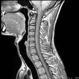
Magnetic resonance imaging (MRI) uses radiofrequency waves and a strong magnetic field rather than x-rays to provide remarkably clear and detailed pictures of internal organs and tissues. The technique has proven very valuable for the diagnosis of a broad range of pathologic conditions in all parts of the body including cancer, heart and vascular disease, stroke, and joint and musculoskeletal disorders. MRI requires specialized equipment and expertise and allows evaluation of some body structures that may not be as visible with other imaging methods.


 What to Expect

MRI Aron Unit

Before the procedure - You will be asked to complete and sign an MRI Safety Screening Form. The technologist will ask you questions related to your health history. Please provide as complete a history as possible. If you have any questions or concerns about the exam, please feel free to ask the technologist before the exam has begun.

To ensure your safety, the technologist may also repeat a number of the items listed on the Screening Form.

You will be asked to remove all metal objects, including all body piercings, before the exam. Purses, billfolds and personal items will be placed in a security box.

Please use the restroom prior to your exam appointment. Once the exam has begun, any interruption may result in restarting the exam.

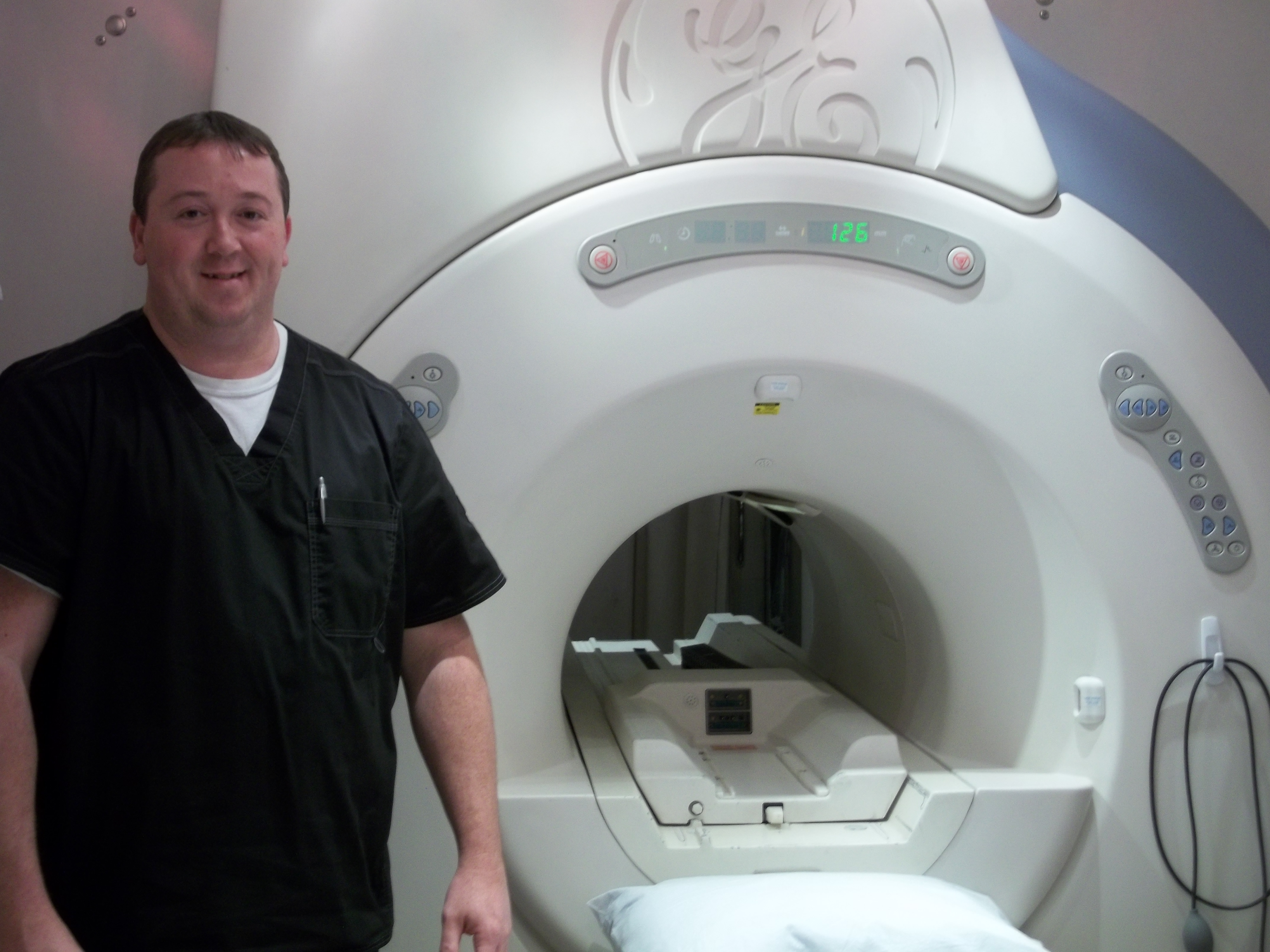
The Procedure - A technologist will position you as comfortably as possible on the scanning table. Depending on the body part to be examined, you will enter the scanner either feet-first or head-first, as the part to be examined must be in the center of the magnet for the machine to work properly. You will be given earplugs to muffle the loud tapping noises of the machine. You may also be given an alert bulb so you may summon the technologist at any time during the exam.

The technologist will perform your exam from an adjoining control room. You will be able to communicate with the technologist with the aid of the alert bulb and an intercom system. It is important that once the technologist positions you for the exam you remain very still and breathe normally. Throughout the exam you will hear loud knocking noises within the machine.

MRI Aron DeskYou may be required to have an injection during the process of the exam. This injection is generally in your arm and consists of a contrast material that will highlight certain structures in your body (the contrast generally has very few adverse effects.) The radiologist and/or your doctor will determine if the contrast material is necessary based on your personal history. Each MRI exam requires several sets of images, called sequences, which may last for several minutes. The average exam will take 30-45 minutes.

After the procedure - You should arrange to get the results of the exam from your doctor, as he/she will receive a report. You should feel no side effects from the scan and may go about your normal daily routine.

How Does It Work?

Unlike conventional x-ray examinations and computed tomography (CT) scans, MRI does not depend on ionizing radiation. Instead, while in the magnet, radio waves redirect alignment of hydrogen atoms that naturally exist within the body without causing any chemical changes in the tissues. As the hydrogen atoms return to their usual alignment, they emit energy that varies according to the type of body tissue in which they lie. The MR scanner listens for this energy and creates a picture of the tissues scanned.

The magnetic field is produced by passing an electric current through wire coils in most MRI units. Other coils, located in the machine and in some cases, placed around the part of the body being imaged, send and receive radio waves, producing signals that are detected by the coils.

A computer then processes the signals and generates a series of images, each of which shows a thin slice of the body. The images can then be studied from different angles by the interpreting radiologist.

Frequently, the differentiation of abnormal (diseased) tissue from normal tissues is better with MRI than with other imaging modalities such as x-ray, CT and ultrasound.

During & After the Procedure

Most MRI exams are painless. However, some patients find it uncomfortable to remain still during MR imaging. Others experience a sense of being closed-in (claustrophobia). Therefore, sedation can be arranged for those patients who anticipate anxiety, but fewer than one in 20 require medication.

It is normal for the area of your body being imaged to feel slightly warm, but if it bothers you, notify the radiologist or technologist. It is important that you remain perfectly still while the images are being obtained, which is typically only a few seconds to a few minutes at a time. You will know when images are being recorded because you will hear and feel loud tapping or thumping sounds when the coils that generate the radiofrequency pulses are activated. Some centers provide earplugs, while others use headphones to reduce the intensity of the sounds made by the MRI machine. You will be able to relax between imaging sequences, but will be asked to maintain your position without movement as much as possible.

You will usually be alone in the exam room during the MRI procedure. However, the technologist will be able to see, hear and speak with you at all times using a two-way intercom. Many MRI centers allow a friend or parent to stay in the room as long as they are also screened for safety in the magnetic environment.

Children will be given appropriately sized earplugs or headphones during the exam. MRI scanners are air-conditioned and well-lit. Music may be played through the headphones to help you pass the time.

In some cases, intravenous injection of contrast material may be performed. The intravenous needle may cause you some discomfort when it is inserted and you may experience some bruising. There is also a very small chance of irritation of your skin at the site of the IV tube insertion. Some patients may sense a temporary metallic taste in their mouth after the contrast injection.

If you have not been sedated, no recovery period is necessary. You may resume your usual activities and normal diet immediately after the exam. On very rare occasions, a few patients experience side effects from the contrast material, including nausea and local pain. Similarly, patients are very rarely allergic to the contrast material and experience hives, itchy eyes or other reactions. If you experience allergic symptoms, notify the technologist. A radiologist or other physician will be available for immediate assistance.